Form Vascular Networks in Microfluidics Model
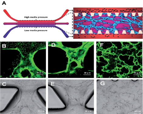
In this study, the authors developed a 3D functional human microvascular network in a microfluidic device. The established model enables Neuromics GFP-labeled human umbilical vein endothelial cellsto form vessel-like microtissues and have physiological functions which are closer to cells in human blood vessels. The perfusable microvasculature allows the delivery of nutrients, and oxygen, as well as flow-induced mechanical stimuli into the luminal space of the endothelium. The microflow effectively mimic the blood flow in human vessels.

This in-vivo like model is then used for toxicity assays-Yan Li, Qing-Meng Pi, Peng-Cheng Wang, Lie-Ju Liu, Zheng-Gang Han,Yang Shao, Ying Zhai, Zheng-Yu Zuo, Zhi-Yong Gong, Xu Yang and Yang Wu. Functional human 3D microvascular networks on a chip to study the procoagulant effects of ambient fine particulate matter. : RSC Adv., 2017, 7, 56108
Our human primary and stem cells are widely used and frequently published. We will continue to post relevant results from researchers using the cells here.
Images: Microvascular network formation based on microfluidic 3D HUVEC culture. (A) Schematic diagram of a microfluidic device. (B) Schematic diagram of microvascular network formation based on microfluidic 3D HUVEC culture. (C) Schematic diagram of loading microparticles in microvascular networks. (D) Microscope image of HUVECs seeding in fibrin hydrogel. (E) Confocal microscope image of fluorescent microvascular networks


