
Browse through our application examples demonstrating, how IBA products were used in the various life science research projects conducted by both our customers and our in-house team of scientists and which results were obtained.
Cell Isolation
- Fab-TACS® Affinity Chromatographic Isolation
CD3+ T cells were isolated from buffy coats or murine splenocytes using CD3 Fab-Streps and Strep-Tactin-TACS® Agarose columns according to the affinity chromatographic Fab-TACS® approach. Purity, yield and viability were evaluated.
Purity
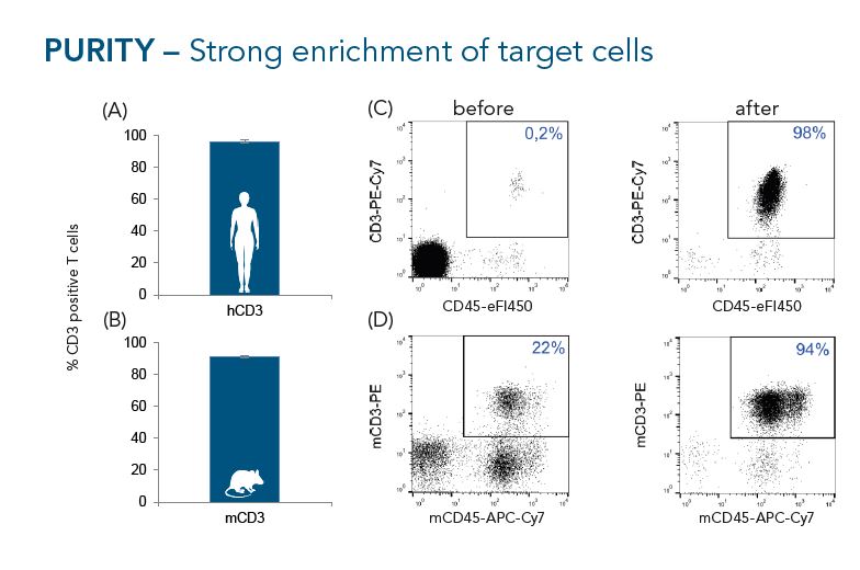
CD3+ cells were isolated from buffy coats (A, n=10) or murine splenocytes (B, n=7) using the Fab-TACS® approach for affinity chromatographic cell isolation. Data are displayed as mean ± SEM. Cells were analyzed by flow cytometry. Representative FACS plots before and after enrichment are shown in C (human) and D (mouse). Single cells were gated according to scatter properties and dead cells excluded by DAPI staining. Isolated human CD3+ T cell had an average purity of 96% and isolated murine CD3+ T cells an average purity of 91%.
Yield
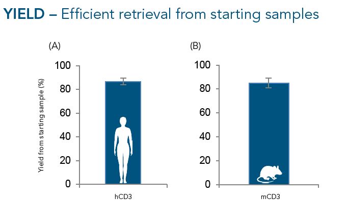
CD3+ cells were isolated from buffy coats (A, n=10) or murine splenocytes (B, n=7) using the Fab-TACS® approach for affinity chromatographic cell isolation. Data are displayed as mean ± SEM. On average, around 85% of CD3+ cells present in the starting material of both human and murine samples were retrieved after CD3-specific enrichment.
Viability
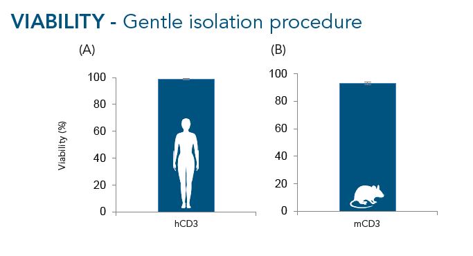
CD3+ cells were isolated from buffy coats (A, n=10) or murine splenocytes (B, n=7) using the Fab-TACS® approach for affinity chromatographic cell isolation. Data are displayed as mean ± SEM. On average, 99% of cells were viable after isolation from buffy coats and 91% after enrichment from murine splenocytes.
- Fab-TACS® Magnetic Isolation
T cells (CD3+, CD4+ and CD8+) were isolated using Fab-Streps and Strep-Tactin® Magnetic Microbeads according to the Fab-TACS® approach for magnetic cell isolation. Purity and viabiliy were evaluated.
Purity
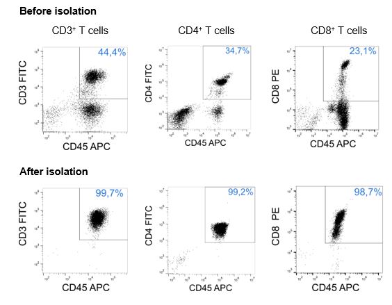
CD3+, CD4+ or CD8+ T cells were isolated from PBMCs using Fab-Streps and Strep-Tactin® Magnetic Microbeads according to the Fab-TACS® approach for magnetic cell isolation. Unlysed cells were either stained with CD3-FITC (OKT-3), CD4-FITC (SK3), or CD8-PE (HIT8a), and CD45-APC (2D1), and analyzed by flow cytometry (CytoFlex, BC). Dead cells were excluded from the analysis using DAPI staining. Doublet and debris discrimination were performed using different FSC/SSC signals. Purity was > 98% for all T cell populations after isolation.
Viability
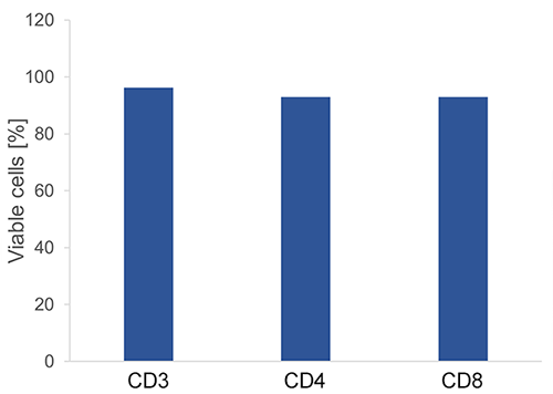
CD3+, CD4+ or CD8+ T cells were isolated from PBMCs using Fab-Streps and Strep-Tactin® Magnetic Microbeads according to the Fab-TACS® approach for magnetic cell isolation. Dead cells were determined using DAPI staining. Doublet and debris discrimination were performed using different FSC/SSC signals. > 93% of cells were viable after all isolation procedures.
Cell Stimulation & Expansion
- CD3/CD28 Streptamer®
CD3+ T cells were isolated from PBMCs using the magnetic Fab-TACS® cell isolation method. Cells were stimulated and expanded with CD3/CD28 Streptamers as indicated. T cell activation, proliferation, fold expansion and cluster formation were evaluated.
Activation
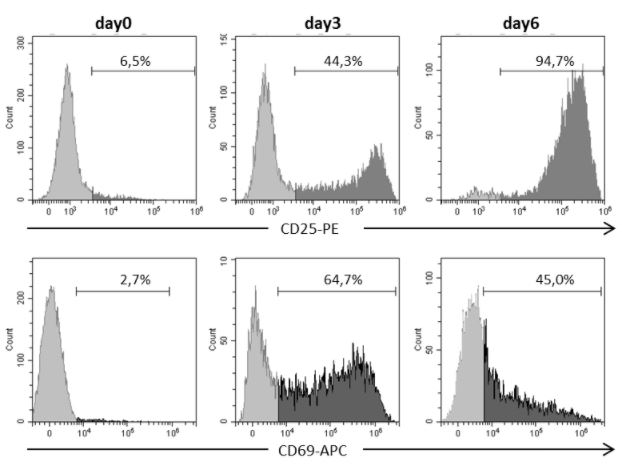
Human CD3+ T cells were isolated with CD3 Fab-Streps and Strep-Tactin® Magnetic Microbeads according to the Fab-TACS® approach for magnetic cell isolation. Activation markers CD25 or CD69 were assessed in pregated CD3+ T cells using Flow Cytometry. Dead cells were excluded from the analysis using DAPI.
Proliferation
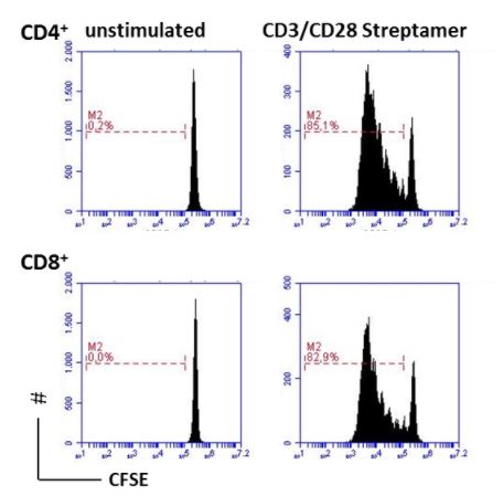
Human CD3+ T cells were isolated with CD3 Fab-Streps and Strep-Tactin® Magnetic Microbeads according to the Fab-TACS® approach for magnetic cell isolation. Cells were stained with CFSE and cultured with CD3/CD28 Streptamers in a 48 well plate for 5 days. Proliferation was measured in pregated CD4+ (upper panel) or CD8+ T cells (lower panel) using Flow Cytometry. Dead cells were excluded from the analysis using PI.
Fold Expansion
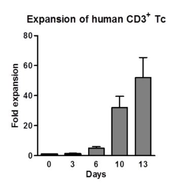
Human CD3+ T cells were isolated with CD3 Fab-Streps and Strep-Tactin® Magnetic Microbeads according to the Fab-TACS® approach for magnetic cell isolation. Cells were cultured for 13 days with CD3/CD28 Streptamers for cell expansion. Fold expansion was measured.
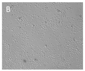
Cluster Formation
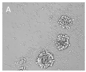
Human CD3+ T cells were isolated with CD3 Fab-Streps and Strep-Tactin® Magnetic Microbeads according to the Fab-TACS® approach for magnetic cell isolation. Cells were cultured with (A) or without (B) CD3/CD28 Streptamers for T cell expansion. Cluster formation was evaluated using microscopy.
Cloning & Transfection
- MEXi - Mammalian Expression System
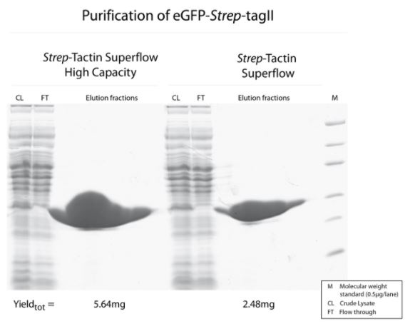
The MEXi system consisting of MEXi 293E cells, pDSG-IBA expression vectors, MEXi-TM transfection medium, and MEXi-CM culture medium was applied for expression of different proteins. Subsequent purification occurred either with Strep-Tactin® Superflow® high capacity or Strep-Tactin®XT Superflow®. Yield and purity were analyzed by SDS-PAGE.
Metalloprotein
Type: metalloprotein, hydrolase
Yield: 143 mg/l.
Secreted Alkaline Phosphatase (SEAP) was fused with a C-terminal Twin-Strep-tag® and the BM40 secretion signal via cloning into pDSG-IBA102. MEXi 293E cells (1.5 x 10⁶ cells/ml) were transfected in MEXi-TM transfection medium (17 ml) with polyethylenimine (PEI, 25 kDa). Afterwards, the cell culture was incubated for 4 hours (37 °C, 5% CO2, 125 rpm) and then diluted with MEXi-CM culture medium to reach a cell density of 0.75 x 10⁶ cells/ml. The cells were kept at 37 °C, 5% CO2, and 125 rpm for 7 days in order to obtain high protein yields. For purification, the cells were pelleted and the supernatant, containing the SEAP protein, was harvested. Free Biotin was blocked by BioLock containing avidin. The SEAP protein was finally purified using a gravity flow Strep-Tactin® Superflow® high capacity column.
Antibody
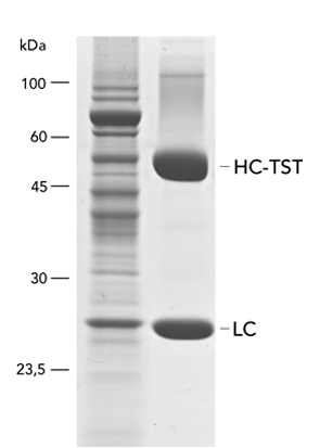
Type: antibody
Yield: 96 mg/l (represented by heavy (HC) and light chain (LC))
The monoclonal rat antibody (mAb) was cloned into the pDSG-IBA102 vector in order to fuse the heavy chain (HC) C-terminally with the Twin-Strep-tag® and the BM40 secretion signal. The transfection of MEXi 293E cells was performed in MEXi-TM transfection medium (250 ml) with polyethylenimine (PEI, 25 kDa). The cells were incubated for 4 hours (37 °C, 5% CO2, 125 rpm) and when a cell density of 3 x 10⁶ cells/ml was reached, 250ml MEXi-CM culture medium was added. Afterwards, the culture was shifted to 32 °C and incubated until day 10.
In order to divide the cells from the supernatant, the cell suspension was centrifuged according to the MEXi manual. Free biotin was blocked by BioLock containing avidin. The supernatant was used for protein purification via a gravity flow Strep-Tactin® Superflow® high capacity column. WET FRED was used to facilitate loading of the large supernatant volume onto the column.
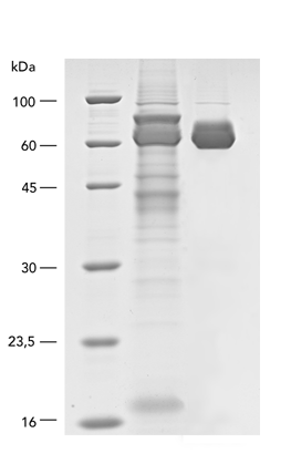
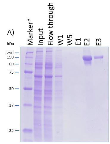
IBA Customer Protein
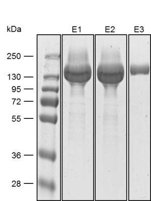
Yield: 318 mg/l
The coding sequence of the customer protein was cloned into pDSG-IBA102 leading to a recombinant protein with C-terminal Twin-Strep-tag® and BM40 secretion signal. 1050 ml of MEXi-TM was inoculated with MEXi 293E cells. Subsequently, plasmid DNA was added followed by addition of 25 kDa linear PEI. The cells were incubated for 4 hours in MEXi-TM medium at 37 °C, 5% CO2, and 125 rpm in an orbital shaking incubator. Cells were diluted to 7.5 x 10⁵ cells/ml by addition of one volume MEXi-CM and kept at 37 °C for 7 days. Afterwards, cells were pelleted and the supernatant containing the customer protein was harvested. The customer protein was finally purified using Strep-Tactin®XT Superflow®. The figure shows the elution fractions (E1-E3). A dilution of 1:10 was prepared for E1 and E2 for SDS-PAGE analysis.
- StarGate Cloning
StarGate comprises vectors for expression in mammalian cells (pCSG-IBA, pESG-IBA, pDSG-IBA), yeast (pYSG-IBA), E. coli (pASG-IBA), and insect cells (pLSG-IBA). As an example, the bacterial protein azurin was cloned into different pASG-IBA vectors and expressed in E. coli. Expression results were analyzed by SDS-PAGE.
pASG-IBA
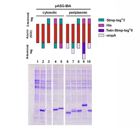
The coding sequence of azurin, a 14 kDa bacterial protein, was cloned into 10 different pASG-IBA vectors and expressed in E. coli. Comparable amounts of E. coli cells were harvested 3 hours after induction with anhydrotetracycline. Samples were lysed by boiling for 5 min at 95 °C with gel loading buffer and analyzed by SDS-PAGE. Subsequently, the gel was stained with Coomassie. Periplasmic secretion by means of ompA led in all cases to accumulation of comparable amounts of the protein of interest (lanes 6-10). This could be expected as azurin is also secreted in its authentic host P. aeruginosa. In case of cytosolic expression, however, interesting aspects became obvious, since it was not anticipated that cytosolic expression of azurin is possible at all. Expression was enabled by fusion of an N-terminal affinity tag (lanes 2, 4, and 5), while N-terminal untagged variants did not result in expression (lane 1 and 3). The examples show that initial screening for expression conditions may be worthwhile since different constructs can lead to various results depending on the target protein properties.
Exosome Isolation
- Fab-TACS® for exosomes
Exosomes were isolated from mesenchymal stem stell supernatants or human serum using Fab-TACS® for exosomes or PEGylation.
Mesenchymal Stem Cells
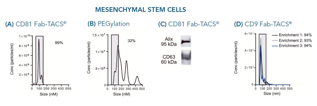
The particle size is a critical factor in evaluating the quality of isolated exosomes. 99% of particles isolated from cell culture supernatants of mesenchymal stem cells (MSCs) using Fab-TACS® for exosomes fell within the range of 30 - 150 nm (A). This indicates strong exosome enrichment. In comparison, only 32% exosome-sized particles were detected after purification with a commercially available PEGylation kit (B), implying contamination by larger extracellular vesicles.
Since other same-sized non-exosome contaminants may contribute to the pool of isolated particles, we tested for the presence of marker proteins CD63 and Alix after exosome isolation from MSC supernatant using our Fab-TACS® approach. Both proteins were clearly present within the purified particles (C), confirming their exosome phenotype.
Depending on MSC donor, cell culture supernatant compositions may vary. Purified exosomes of three independent isolations from different MSC donors were very comparable in their size ranging from 86 nm to 90 nm average diameter. All isolations yielded around 94% of particles between 30 and 150 nm in size (D).
Human serum
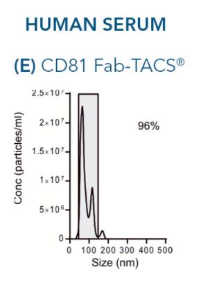
Important exosome sources also include a variety of human bodily fluids such as blood and urine. Similar to isolations from cell culture supernatants, 96% of particles retrieved by exosome isolation from serum of humans exhibited the typical exosome size of 30 to 150 nm (E).
Protein Affinity Chromatography
- Protein purification with Strep-Tactin®XT
For protein purification of Strep-tag®II and Twin-Strep-tag® proteins with Strep-Tactin®XT IBA Lifesciences offers MagStrep "type 3" XT beads , 4Flow®, and pre-packed Spin Columns. As example, the purification of different target proteins with Strep-Tactin®XT 4Flow® and MagStrep "type 3" XT beads is shown.
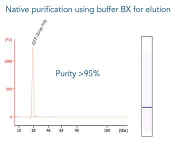
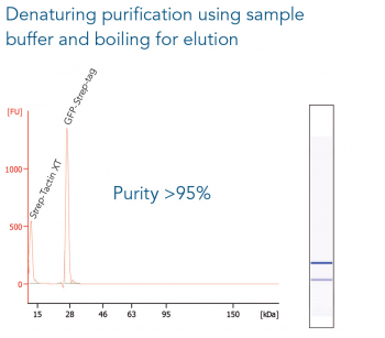
MagStrep “type3” XT beads
GFP C-terminally tagged with Strep-tag®II (28 kDa) was expressed in E. coli (A) and purified with MagStrep “type3” XT beads. For separation of magnetic beads from residual solution, the Magnetic Separator was used. Before elution, the sample was split and the target protein was eluted either by application of 1x Buffer BXT containing biotin or by boiling (C, 5 min at 95 °C). Due to boiling, the Beat´s agarose melts and Strep-Tactin®XT is released, leading to a further peak at 14 kDa. Protein purification results were analyzed with the Agilent Bioanalyzer 2100 system. The example shows the specific binding properties of MagStrep “type3” XT beads and the high purity that can thereby be observed.

Strep-Tactin®XT 4Flow®
Cell culture supernatant of MEXi-293E cells (24 ml) was spiked with 240 µg of a large protein (130 kDa) C-terminally tagged with the Twin-Strep-tag®. Protein purification was carried out with a 0.2 ml Strep-Tactin®XT 4Flow® high capacity gravity flow column. After sample application the column was washed with 1x Buffer W. The Twin-Strep-tag® protein was eluted by three elution steps with 0.7, 1.5 and 0.8 CV 1x Buffer BXT. Purification results were analyzed by SDS-PAGE (A) and western blot (B). Both show a molecular weight marker (M*, Precision Plus Protein™ Unstained Protein Standards, Strep-tagged recombinant (BioRad), M** PageRuler™ Plus Prestained Protein Ladder (Thermo Fisher Scientific)), the spiked cell supernatant (S), flow through (FT), the 1st and 5th wash fraction (W1 and W5, respectively) and the elution fractions (E1-E3). Strep-Tactin® HRP was applied (1:4000) for detection via western blot.

- Protein purification with Strep-Tactin®
IBA offers MacroPrep®, Superflow®, and Sepharose® coupled with Strep-Tactin® for protein purification of Strep-tag®II and Twin-Strep-tag® proteins. As example, the purification of different target proteins with Strep-Tactin® Sepharose® or Strep-Tactin® Superflow® is shown.
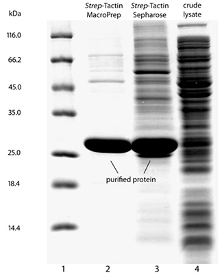
Sepharose®
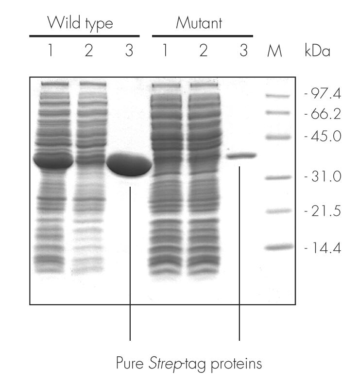
The wild type sequence and mutant variant of an enzyme were cloned into pASK-IBA3, leading to proteins localized to the cytoplasm and C-terminally tagged with the Strep-tag®II. After expression in E. coli and cell lysis, the proteins were purified with Strep-Tactin® Sepharose® gravity flow columns under physiological conditions (100 mM Tris-Cl, pH 8.0). Purification results were analyzed by SDS-PAGE. A molecular weight marker (M), cell lysate (lane 1), flow through (lane 2), and elution with 2.5 mM desthiobiotin (lane 3) are shown. For both proteins, a high purity was observed.
Superflow®
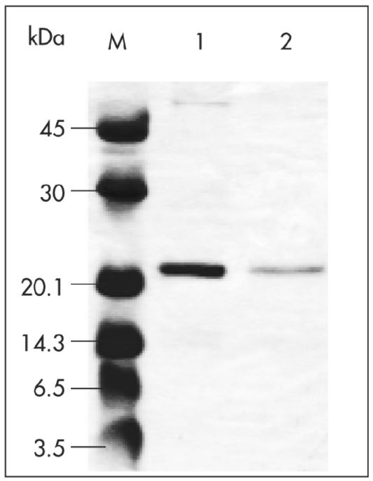
Coding sequences of virus-like particles (VLPs, 4.8 MDa) were cloned into pASK-IBA7 to obtain proteins localized to the cytoplasm and N-terminally tagged with Strep-tag®II. The plasmid was transformed into E. coli and the cell culture (1 l) was induced at OD600 of 0.6 by addition of anhydrotetracycline (AHT). Protein expression was performed at 37 °C for 3 h at 200 rpm. Afterwards, cells were harvested, resuspended with 20 ml 1x Buffer W (100 mM Tris-Cl, 150 mM NaCl, 1 mM EDTA), and sonicated. The insoluble material was pelleted, and the lysate was applied to a Strep-Tactin® Superflow® cartridge (1 ml/min flow rate). After washing, the target protein was eluted with 2.5 mM desthiobiotin. Purification results were analyzed by SDS-PAGE. Marker (M) and two different elution fractions of VLPs fused to Strep-tag®II (lane 1 and 2) are shown.
Data kindly provided by L. Stöckl and B. Brandenburg, Robert-Koch-Institute, Berlin. Brandenburg, et al. (2005): A Novel System for Efficient Gene Transfer Into Primary Human Hepatocytes Via Cell-Permeable Hepatitis B Virus–like Particle, HEPATOLOGY (42), 1300-1309.
- Resin Comparison
Both Strep-Tactin® and Strep-Tactin®XT resins can be used for purification of Strep-tag®II and Twin-Strep-tag® proteins. However, due to their different properties, an initial comparison can be helpful to find the appropriate resin.
Superflow® vs. Superflow® high capacity
Cell lysate contaning GFP-Twin-Strep-tag was split and either purified with Strep-Tactin® Superflow® or Strep-Tactin® Superflow® high capacity.
Both purifications led to a highly pure protein, but due to the higher density of immobilized Strep-Tactin® on the high-capacity variant, a two-fold higher amount of GFP-Twin-Strep-tag can be purified with Strep-Tactin® Superflow® high capacity compared to Strep-Tactin® Superflow®.
Strep-Tactin® vs. Strep-Tactin®XT
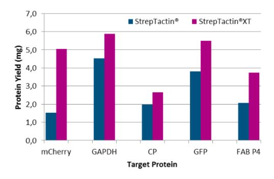
Five different proteins were purified with Strep-Tactin® Superflow® and Strep-Tactin®XT Superflow® to compare the yield with both rein types. Here, we found that Strep-Tactin®XT Superflow® allows the purification of on average a 2-fold higher protein yield than Strep-Tactin® Superflow®.
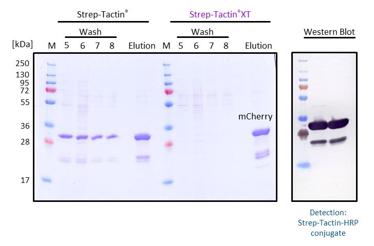
An advantage of Strep-Tactin®XT compared to Strep-Tactin® is that it allows intensive washing without loss in protein yield. To illustrate this, mCherry-Twin-Strep-tag from E. coli supernatant was purified with a gravity flow column either containing 1 ml Strep-Tactin® Superflow® or Strep-Tactin®XT Superflow®. During purification, the columns were washed with 8 column volumes of 1x Buffer W to reach a constant absorption ratio at A260/280 nm. Then, the target protein was eluted with 50 mM biotin. Samples of the last wash fractions (wash 5-8) and the elution fraction were analyzed by SDS-PAGE. In comparison to Strep-Tactin® Superflow®, Strep-Tactin®XT Superflow allows intensive washing without loss of protein yield. The high affinity (pM range) between Strep-Tactin®XT and Twin-Strep-tag® leads to a tighter binding and prevents a premature elution of the target protein during the wash steps. The elution fractions were further analyzed by Western Blot and detected with Strep-Tactin®HRP, showing that the further bands are alternative variants of mCherry-Twin-Strep-tag.
MacroPrep® vs. Sepharose®
Cell lysate contaning GFP-Twin-Strep-tag was split and either purified with Strep-Tactin® Superflow® or Strep-Tactin® Superflow® high capacity.
Both purifications led to a highly pure protein, but due to the higher density of immobilized Strep-Tactin® on the high-capacity variant, a two-fold higher amount of GFP-Twin-Strep-tag can be purified with Strep-Tactin® Superflow® high capacity compared to Strep-Tactin® Superflow®.
- Antibody purification with Protein A
Protein A allows the the tag-free purification of IgG class antibodies. Dynamic binding capacity and stability were evaluated.
Dynamic Binding Capacity
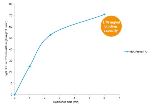
Dynamic binding capacity (DBC 10%) of IBA Protein A determined with a monoclonal antibody.
Per milliliter Protein A resin more than 70 mg of the monoclonal antibody can be captured and purified.
Stability
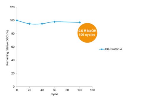
Dynamic binding capacity of Protein A remains stable after treatment with 0.5 M NaOH over~100 cycles (1 cycle = 15 min).
Protein-Protein Interaction
- Strep-Tactin®XT
Strep-Tactin®XT binds with high affinity Twin-Strep-tag® proteins, making it perfectly suitable for SPR analysis. The examples show the coupling of Strep-Tactin®XT on different SPR chips and possible regeneration conditions for re-use of Strep-Tactin®XT-coated chips.
Coupling on CM5
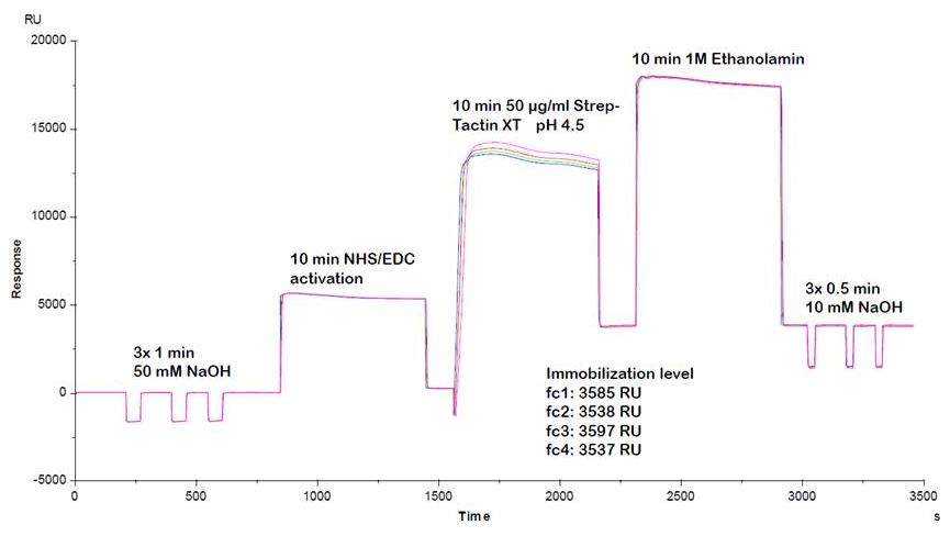
We recommend to use the following conditions for optimal Strep-Tactin®XT coupling:
- 20-50 µg/ml Strep-Tactin®XT
- max. 20 mM acetate, pH 4.5
Using these conditions for Strep-Tactin®XT coupling, efficient and reproducible coupling results can be obtained.
Coupling on CM4
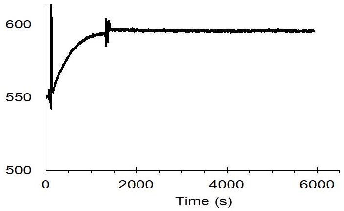
The membrane protein CB2 was fused with Twin-Strep-tag® and then immobilized on a CM4 sensor chip coated with Strep-Tactin®XT.
Used conditions:
- 50 mM Tris pH 7.5, 100 mM NaCl
- 10 % glycerol, 0.1 % DDM, 0.5 % CHAPS, 0.1 % CHS
Data was published in A. Yeliseev et al., Protein Expression and Purification 131 (2017) 109-118
Regeneration
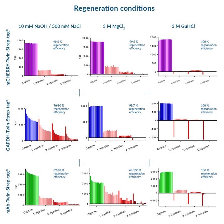
Three different regeneration conditions were tested on Strep-Tactin®XT CM5 sensorchips:
- 10 mM NaOH / 500 mM NaCl
- 3 M MgCl2
- 3 M GuHCl
for three different Twin-Strep-tag® fusion proteins:
- mCherry-Twin-Strep-tag
- GAPDH-Twin-Strep-tag
- mAb-Twin-Strep-tag
We recommend the application of 3 M GuHCl for optimal regeneration conditions, since this buffer led to the best regeneration results for all test proteins. However, depending on the target protein, other buffers are applicable as well.
- StrepMAB-Immo
Due to the nearly irreversible binding of Strep-tag®II proteins, StrepMAB-Immo can be used for SPR analysis. The example shows the stable capture of a Strep-tag®II fusion protein on a SPR chip coated with StrepMAB-Immo.
Surface Plasmon Resonance
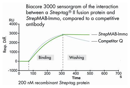
Chips for SPR were coated either with StrepMAB-Immo or with a competitive antibody (Competitor Q). Subsequently, a Strep-tag®II fusion protein was captured, and the binding stability was determined using a Biacore 3000.
During the washing phase, the recombinant Strep-tag® protein of interest remains tightly bound to StrepMAB-Immo, while a significant amount of Strep-tag® protein is washed off using the competitive antibody.
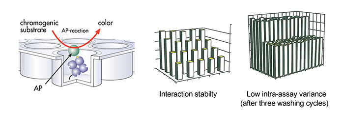
- Strep-Tactin® microplates
Strep-tag®II fusion proteins were immobilized on Strep-Tactin® microplates. Subsequently, the activity or abundance of the Strep-tag®II fusion proteins was determined by enzymatic reaction.
Activity Assay
Strep-Tactin® coated microplate was incubated with different amounts of recombinant E. coli alkaline phosphatase (AP) tagged with the Strep-tag®II. After several washing steps, activity of bound fusion protein was determined by colorimetric reaction.
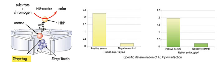
Solid phase immunoasay
Strep-Tactin® coated microplate was incubated with recombinant H. pylori urease tagged with Strep-tag® II, followed by three washing cycles. Afterwards, the microplate was incubated with human sera followed by three washing cycles. Third incubation occurred with rabbit anti-human lgG conjugated to horseradish peroxidase (HRP), followed by 3 washing cycles. Amount of bound antibodies from human sera was determined by colorimetric reaction of HRP.
Staining & Detection
- Protein detection with Strep-Tactin® Conjugates
Strep-Tactin® conjugates can be used for several assay applications. As example, the application of Strep-Tactin®HRP and Strep-Tactin®AP for Western Blot as well as Dot Blot is shown.
Strep-Tactin® HRP
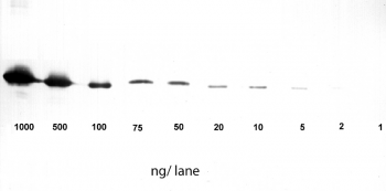
Application: Western Blot
1-1000 ng/lane of GFP C-terminally tagged with Strep-tag®II (28 kDa) were separated by SDS-PAGE and blotted onto a membrane. Detection occurred with Strep-Tactin® HRP.
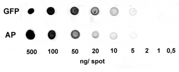
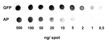
Application: Dot Blot
0.5-500 ng/spot of GFP-Strep-tagII (28 kDa) or alkaline phosphatase C-terminally tagged with Strep-tag®II (monomer 48.5 kDa) were spotted onto a membrane. Detection occurred with Strep-Tactin® HRP.
Strep-Tactin® AP
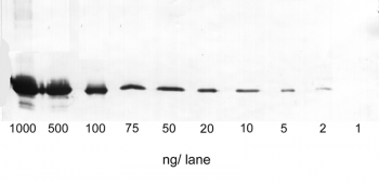
Application: Western Blot
1-1000 ng/lane of GFP C-terminally tagged with Strep-tag®II (28 kDa) were separated by SDS-PAGE and blotted onto a membrane. Detection occurred with Strep-Tactin® AP.
Application: Dot Blot
0.5-500 ng/spot of GFP-Strep-tagII (28 kDa) or alkaline phosphatase C-terminally tagged with Strep-tag®II (monomer 48.5 kDa) were spotted onto a membrane. Detection occurred with Strep-Tactin® AP.
- Detection with Strep-tag® Antibodies
StrepMAB-Classic and StrepMAB-Immo can be used for detection of Strep-tag®II or Twin-Strep-tag® proteins on cell surfaces, within the cell, in cell lysates, or in eluates after protein purification. As example, the application for flow cytometry and confocal microscopy is shown.
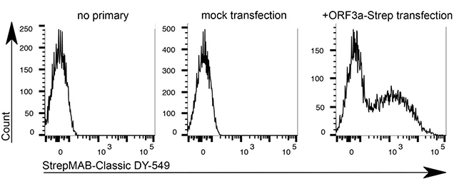
StrepMAB-Classic DY-549
Application: Flow cytometry
Dilution: 1:500
HeLa cells were transiently transfected with the coding sequence of SARS-CoV-2 ORF3a-Twin-Strep-tag. After 48 hours transfection, cells were detached, gently trypsinised and stained with StrepMAB-Classic DY-549 (1:500, 0.5% BSA + 1 mM EDTA, 30 min on ice). Intracellular SARS-CoV-2 ORF3a-Twin-Strep-tag levels were measured by flow cytometry (4 laser Cytoflex S, Beckman Coulter)
Source: Data kindly provided from James Edgar, Department of Pathology at the University of Cambridge.
StrepMAB-Classic
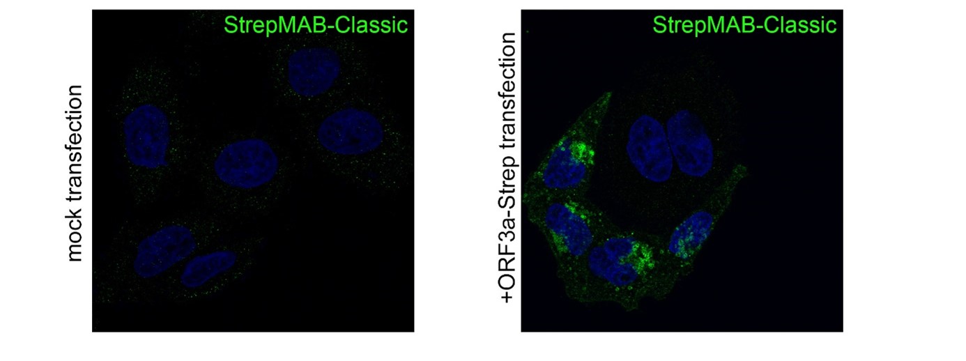
Application: Confocal microscopy (Immunofluorescence)
Dilution: 1:500
HeLa cells were grown on glass coverslips, fixed (4% PFA/PBS), quenched (15 mM glycine/PBS), permeabilized (0.1% saponin/PBS), blocked (1% BSA, 0.01% saponin in PBS), and stained with StrepMAB-Classic (1:500) to confirm the expression of SARS-CoV-2 ORF3a-Twin-Strep-tag. Anti-mouse 488 was used as secondary antibody. Nuclei were stained with DAPI and the imaging occurred with a LSM700 confocal microscope (63×/1.4 NA oil immersion objective; ZEISS).
Source: Data kindly provided from James Edgar, Department of Pathology at the University of Cambridge.
- MHC I Streptamer® Cell Staining
Different antigen-specific T cells were stained with MHC I-Streps and fluorescent Strep-Tactin® conjugates. All experiments were performed in close cooperation with D. Busch, TU Munich.
CMV-specific CD8+ T cells
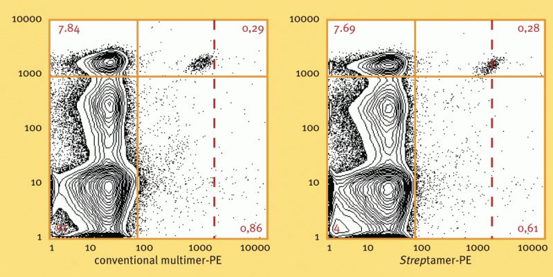
Ex vivo staining of CMV-specific CD8+ T cells with HLA-A*0201 CMV pp65495-503 Streptamer® (right diagram) or conventional multimers (left diagram). Staining was performed from a CMV positive individual at 4°C. Staining with MHC I Streptamer® resulted in improved staining intensity.
Used reagents:
- 6-7001-001 MHC I-Strep CMV, pp65495-503
- 6-5000-001 Strep-Tactin® PE
LLO-specific CD8+ T cells
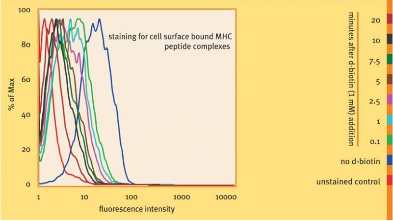
Antigen-specific cells were visualized with Strep-Tactin® PE coupled to MHC I-Strep H2-Kd specific for LLO91-99 . After addition of 1 mM d-biotin, subsequent dissociation of monomerized MHC molecules was monitored at different time points as indicated. 20 minutes after biotin addition, staining reagents were largely removed from the cells. Staining and dissociation were performed at 4 °C.
Used reagents:
- 6-7014-001 MHC I-Strep H2-Kd, LLO91-99
- 6-5000-001 Strep-Tactin® PE
Her2/neu-specific CD8+ T cells
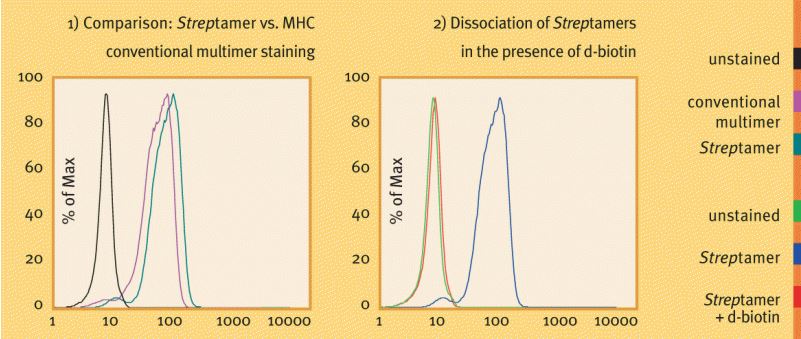
Staining of Her2/neu-specifc CD8+ T cells (kindly provided by H. Bernhard, TU Munich) with HLA-A*0201/Her2/neu369-377 Streptamers® as compared to conventional multimers (left diagram). Complete dissociation of Streptamer® staining reagents from the cells is shown after a brief incubation (30 min) in the presence of 1 mM d-biotin (right diagram). Antigen-specific T cells were visualized with Strep-Tactin® PE coupled to MHC I-Strep HLA-A*0201 specific for Her2/neu369-377. Staining and dissociation were performed at 4 °C.
Used reagents:
- 6-7008-001 MHC I-Strep HLA-A*0201, Her2/neu369-377
- 6-5000-001 Strep-Tactin® PE