In the past couple of weeks, our blog has shared studies using Neuromics GFP-expressing cells and cell lines in exciting research. Now, we are happy to highlight another new publication that merges the two—a GFP-expressing cell line. Last month, researchers at Dartmouth published a paper using our GFP-expressing human glioblastoma cells (U87 MG) (cat. TR01-GFP) in fluorescent-guided surgery research.
Contrast imaging has become a frequently used tool in surgical resection so that surgeons can remove all tumor tissue when operating on patients. This method has especially become prevalent in glioma removal. While many methods for fluorescent-guidance surgery exist, the investigators wanted to look for one that accurately marks tumors shortly after administration and is compatible with current imaging capabilities.
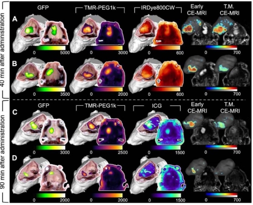
Image: Imaging of mice inoculated with GFP expressing U87 MG cells after 40 and 90 minutes. A comparison with GFP shows how candidate fluorescent agents perform.

The scientists cultured Neuromics GFP-expressing U87 MG cells and then inserted them into a mouse model. After testing several fluorescent agents, they found one, tetramethylrhodamine conjugated to a small polyethylene glycol chain (TMR-PEG1k), performed the best. You can read the complete study here.
Please check out our primary cell and cell line options. All are 15% off through the end of 2024.


